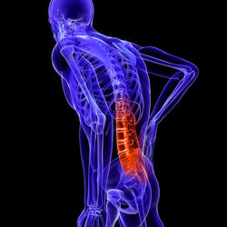
Injury Of The Spinal Column
Thoracolumbar spinal injuries can be subdivided into those involving the thoracic spine from T1 to T10, those involving the thoracolumbar junction from T11 to L2 and those involving the lumbar spine from L3 to L5.
The understanding of pathomechanics and classification will guide the surgeon during assessment and in establishing treatment priorities.
Pathomechanics
 The spine seems particularly vulnerable at the junctional zones to failure during the application of high energy particularly in sport at the thoracolumbar junction. There is a transition from the relatively rigid thoracic spine splinted very effectively by the ribs, sternum and dense ligamentous structures, to the much more flexible lumbar spine where up to 503 of flexion-extension movement and in extension, significant lateral flexion and some axial rotation occurs.
Biomechanically it has proved most simple to consider the spine as anterior and posterior columns separated by the posterior longitudinal ligament. The previous interest in a ‘middle column’ has diminished with the biomechanical revelation that the middle column makes a very minor contribution to spinal stability and is unhelpful in clinical use. The anterior column is predominantly a compression structure and the vertebral bodies and discs are both well suited for sustaining prolonged axial loading in the upright posture and brief peaks of force and pressure during jumping, lifting-type activities.
The posterior column,which is predominantly softtissue, can best be considered as a tension band in all areas of the spine. It consists of a tension element, including the supraspinous and interspinous ligaments attached to the spinous processes, and the bony elements of laminae and facet joint which have multiple biomechanical functions. The facet joints take significant axial loads, particularly in extension,are important components resisting rotational and shear stresses and facilitate movement at each motion segment in the thoracolumbar spine.
The tension band elements of the posterior column are particularly prone to failure during distraction forces, although less commonly distraction forces can injure the anterior column such as in the Chance fracture, commonly associated with disc disruption.
More complex injuries or combination injuries occur with higher energy and with the addition of rotational forces, and this pattern of injury is highly associated with underlying spinal cord injury. These injuries occur with violence greater than usually occurs during contact sports and are seen in high-energy sports such as motorracing and equestrianism.
Instability Instability is a widely misused word that has been defined by White and Panjabi as ‘the loss of the ability of the spine under physiological loads to maintain its pattern ofdisplacement so that there is no initial or additional neurological deficit, no major deformity and no incapacitating pain’. There are undoubtedly clinical, biomechanical and radiological criteria for diagnosing instability but in the clinical situation, if missed, it can produce or exacerbate an underlying spinal cord injury or increase the severity of the pattern of fracture or dislocation.
Certain injuries, particularly the combination of an anterior compression injury with a posterior distraction injury lead to chronic instability and loss of spinal sagittal alignment, and in the long term this will lead to pain and gradual neurological deficit.
Classification
The system of classification of thoracolumbar injuries remains controversial. In the recent past, the Denis three-column mechanistic classification has probably been the most popular system. It is, however, quite cumbersome in clinical use and biomechanical studies suggest that the ‘middle column’ is not really important. This has led to a re-emergence of classification systems based on two-column theories of spinal function, and since it has been adopted by the AO Group and the Orthopaedic Trauma
Association of North America, the Magerl classification is the most useful and widely accepted in current clinical practice. There are three basic injury patterns and these are further sub-divided at several levels; these patterns are:
- Type A compression injuries of the vertebral body
- Type B distraction injuries of anterior or posterior elements
- Type C : Type A or type B injuries with rotation,and complex fractures and dislocations
In its simplest form,the classification is a useful description of injury mechanism based on clinical and radiological assessment and it is very helpful in determining appropriate treatment options. At a more complex level, it is a useful and moderately well reproducible research and audit tool allowing comparison of Pedicle screw reduction and fixation in a thoracolumbar dislocation. the management of different fracture patterns in different centres.
Assessment
The assessment of any potentially injured spine,either at the scene of the sporting accident or in the receiving hospital, should comply with the standard immobilization and resuscitation protocols that are very well addressed in the Advanced Trauma Life Support Course manuals.
The assessing doctor should be especially alert for associated non-spinal injuries and also for a second or even third spinal injury to patients in whom one injury has already been identified. The incidence of a second injury in the spinal column is reported to be as high as 20% in some series.
The orthopaedic assessment of the spine should be focussed on the skeletal and the neurological injuries. A subtle history of transient neurological symptoms may be of critical importance in any spinal injury and careful neurological assessment of all sensory and motor modalities in upper and lower limbs is essential. In the thoracolumbar spine,information about injuries to the posterior column may only be gleaned from clinical assessment. Careful inspection and palpation of the spinal column is important.
Pain on movement of the spine,localized tenderness,or a palpable gap or step should all indicate significant injury to the underlying posterior column structures. After transfer to an appropriate hospital environment,radiological evaluation should include AP and lateral plain X-rays: these are particularly useful for assessing compression injuries of the anterior columns although careful inspection for interspinous widening or pedicle spread may also identify posterior column injuries.
Where any significant spinal injury is identified, CT scanning is mandatory and this provides further information about the fracture pattern in the anterior column. This is the most reliable way of identifying fractures in the posterior column and facet area and two-dimensional reconstructions will frequently reveal interspinous widening or spinous process fractures where the posterior tension band has been disrupted.
Modern MRI sequences are helpful in identifying posterior soft-tissue disruption and may provide useful information about the pathophysiology of neurological injuries. In the thoracolumbar spine dynamic X-rays are generally not indicated in the trauma situation, but if a question exists about the stability of the spinal column following a full radiological and clinical work-up,then sometimes an appropriate erect X-ray in a brace may help in the surgical decision-making process.
Treatment
The majority of neurologically intact patients with spinal column fractures can be treated non-operatively. There is no biomechanical rationale for a brace, particularly at the thoracolumbar junction and in the lumbar spinewhere axial compressive forces cannot be resisted by external splintage. Nonetheless, most sportsmen have a very high level of motivation and often return to a inappropriately high level of physical activity during the healing phase of a spinal column injury. For this reason, the treating physician may use a brace as an activity restraint and both three-point braces and TLSOs (thoracolumbosacral orthoses) have proved useful in this context. A period of up to 3 months should be assumed for full bone healing and activity restriction and external bracing, if used, should be continued during this period of time. In injuries where pain prevents early mobilization, or in significant two-column injuries or other identified instability patterns, surgical stabilization may be considered appropriate. The sportsman will need to be involved in the decision, as most surgical techniques will involve fusion of at least two spinal motion segments and this is likely to lead to fixed, permanent functional deficit that may prevent the sportsman from returning to the level of function enjoyed before surgery.
In severe twocolumn disruption with major deformity there may be little choice but there are some marginally unstable injuries that seem increasingly to be treated surgically,more often for expediency and reduced healthcare cost than for proven clinical benefits. Modern spinal surgical techniques allow safe, strong stabilization of anterior or posterior columns of the spine or both and most such techniques will allow early mobilization of the patient.
Rehabilitation and return to sport Any patient with a spinal fracture,and particularly highly trained and fit athletes, will start to become deconditioned from the moment of the accident. Paravertebral, trunk and anterior abdominal muscles will lose strength and proprioceptive control; these changes will progress during the treatment period. This inevitably leads to vulnerability of the patient to further injury after the initial treatment phase.
Trunk stabilization exercises can commence very early after the injury without any requirement for movement of the spine. After the treatment phase, the patient should enter into a more vigorous, physiotherapist-supervised, trunk stabilization programme using a series of graded exercises.
The sportsman should also commence some low-level aerobic activity such as swimming or running on a treadmill. With progressive reconditioning and under continued supervision, both by the physiotherapist and the trainer, the sportsman should be allowed to find their own pace of return to sports-specific exercise and ultimately back into the sporting environment. Too much haste will probably lead to repeated setbacks and frustrations,and may well jeopardize the sportsman’s career. A properly paced return with adequate anatomical and physiological education by the supervising physiotherapist, doctor ,and coach or trainer experienced in althletes returning to sports after injury is most likely to result in a return to the pre-injury sport.
Realistically, with a significant spinal injury, this will not occur within 1 year of injury. Although difficult to measure, it seems unlikely that spinal function will ever return to 100% following a significant spinal column injury treated either non-operatively or surgically. It is important to introduce this principle to the sportsman early after the injury so that appropriate goals can be set.